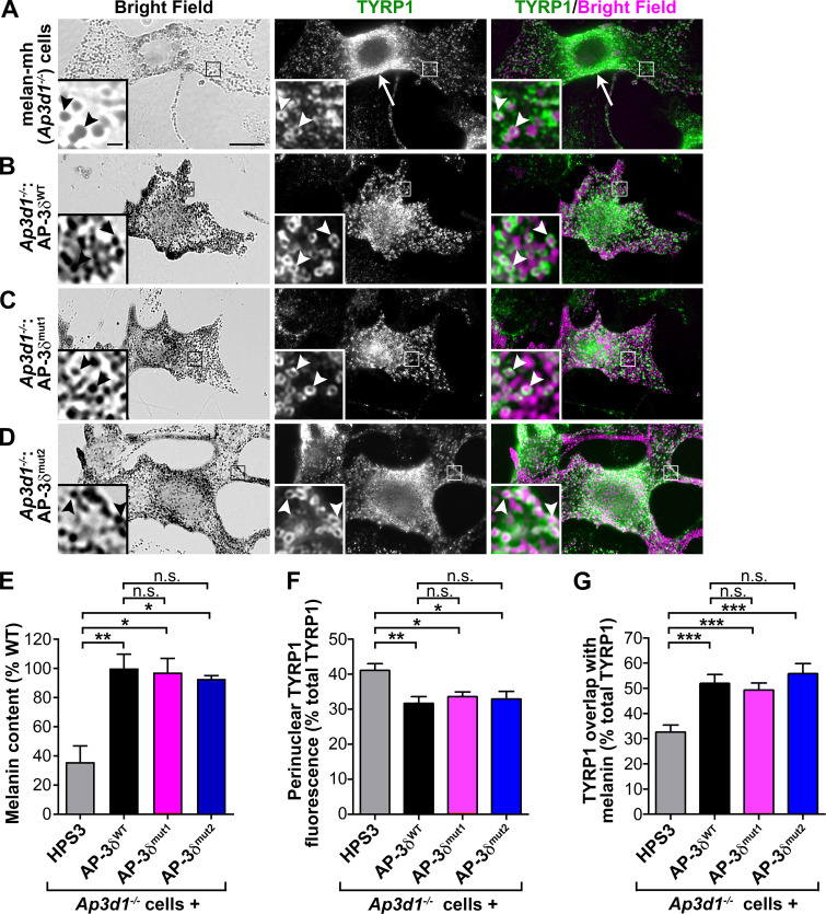
Figure 1.
VAMP7 binding by AP-3δ is not required for normal pigmentation or cargo localization in melanocytes. (A–D) Untransduced (Ap3d1−/−; A) or retrovirally transduced AP-3δ–deficient melan-mh cells stably expressing WT AP-3δ (AP-3δWT; B) or AP-3δ VAMP7 binding mutants (AP-3δmut1, AP-3δmut2; C and D) were fixed, labeled for TYRP1 and STX13, and analyzed by dIFM and brightfield microscopy. Brightfield images (taken at same gain and exposure) are pseudocolored magenta in the right panels. Boxed regions are magnified fivefold in insets. Arrow, TYRP1 accumulation in the perinuclear region of an untransduced cell; arrowheads, TYRP1 on pigmented melanosomes. Scale bar, 5 µm; inset scale bar, 1 µm. (E) Melanin content in cell lysates quantified by spectrophotometry. Data (mean ± SEM from three separate experiments) are normalized to values from melan-mh cells expressing AP-3δWT. (F and G) Quantification of imaging data; melan-mh cells stably expressing HPS3 are the virally transduced negative control. (F) Quantification of TYRP1 perinuclear signal by dIFM relative to total cellular TYRP1 (percent). Data represent mean ± SEM from 25 (HPS3 control), 20 (AP-3δWT), 26 (AP-3δmut1), and 21 (AP-3δmut2) cells over three separate experiments. (G) Quantification of percentage overlap of TYRP1 by dIFM with melanin by brightfield microscopy. Data represent mean ± SEM from 28 (HPS3 control), 20 (AP-3δWT), 34 (AP-3δmut1), and 20 (AP-3δmut2) cells over three separate experiments. *, P < 0.05; **, P < 0.01; ***, P < 0.001.

