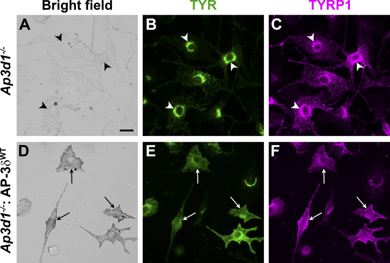
Figure S1.
Disruption of TYR and TYRP1 localization in Ap3d1−/− melan-mh cells. (A–F) Melan-mh cells that were untransduced (A–C) or stably transduced to express WT AP-3δ (melan-mh: AP-3δWT; D–F) were fixed, immunolabeled for TYR and for TYRP1, and analyzed by dIFM and brightfield microscopy. Arrowheads, perinuclear accumulation of both TYR and TYRP1 in hypopigmented melan-mh cells; arrows, peripheral localization of TYR and TYRP1 in darkly pigmented melan-mh: AP-3δWT cells. Scale bar, 20 µm.

