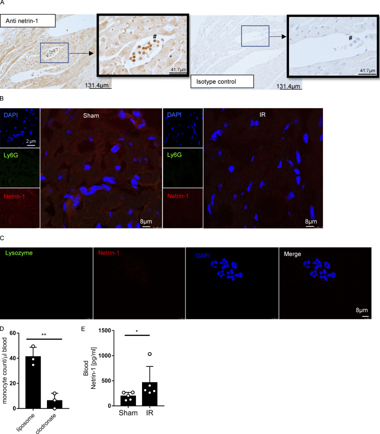
Figure S2.
Netrin-1 staining of heart tissue, negative controls of immunofluorecence, and blood netrin-1 levels of monocyte/macrophage-depleted mice. (A) Immunohistochemical staining for cardiac netrin-1 in WT mice after IR surgery shows positive staining in cardiomyocytes and neutrophils (n = 3; *, cardiomyocytes; #, neutrophils; for original pictures, scale bar = 131.8 µm; for enlarged pictures, scale bar = 41.7 µm). (B) Negative controls for Fig. 1 G (for single-channel pictures, scale bar = 2 µm; for merged channel pictures, scale bar = 8 µm). (C) Negative controls for Fig. 3 H. Scale bar = 10 µm. (D) Murine blood monocyte cell counts after clodronate depletion compared with liposome control group (n = 3 for each group; two-tailed unpaired t test). (E) Blood netrin-1 levels of WT (C57BL/6) mice with clodronate monocyte/macrophage depletion after IR surgery compared with sham group (n = 5 for each group; Mann-Whitney test). *, P < 0.05; **, P < 0.01. Data are presented as mean ± SD.

