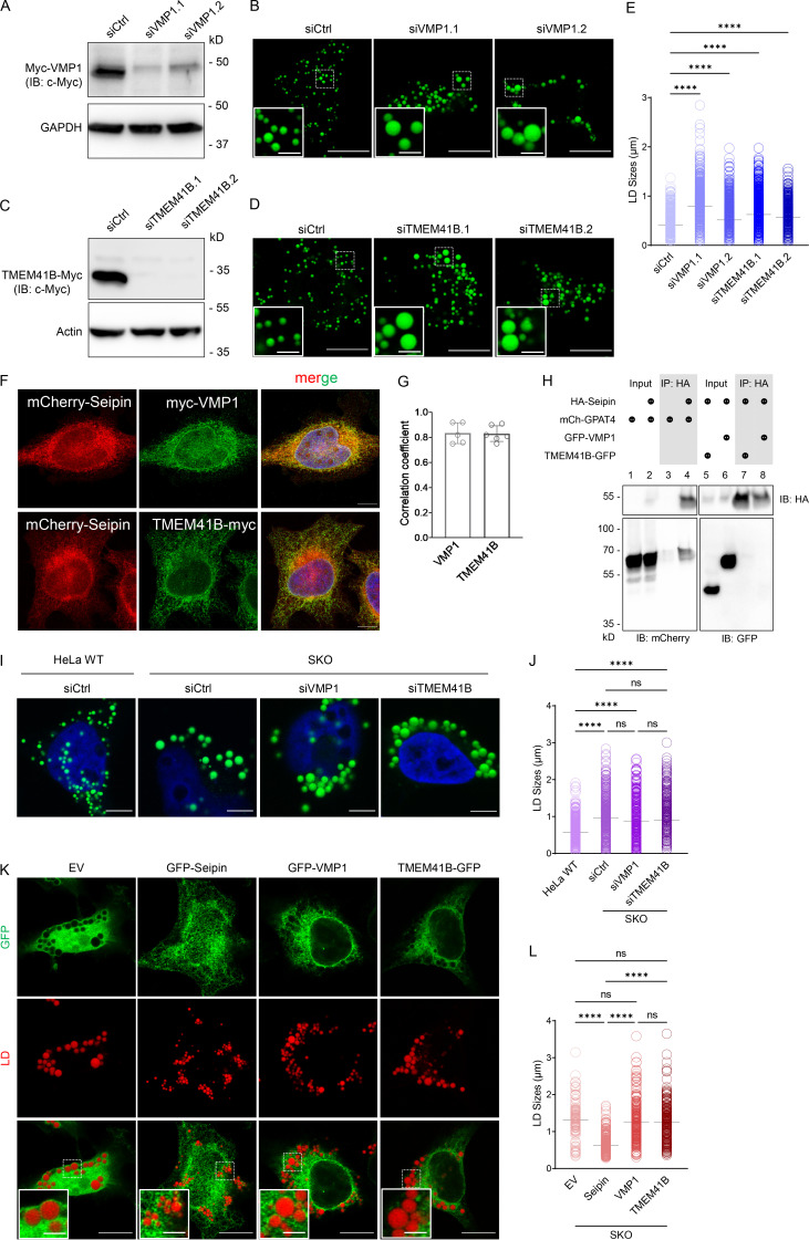
Figure 1.
Seipin and VMP1/TMEM41B regulate LD growth likely through distinct mechanisms. (A and C) Western blot analysis of myc-VMP1 or TMEM41B-myc in HeLa cells treated with control or two different VMP1/TMEM41B siRNAs. Cells were treated with siRNA for 24 h followed by the transfection of myc-VMP1 or TMEM41B-myc cDNA for 24 h. GAPDH/actin was included as loading control. (B and D) Confocal images of oleate-treated (6 h) cells transfected with siRNAs as in A or C and stained with BODIPY. (E) LD diameters measured from images shown in B and D. ****, P < 0.0001 (one-way ANOVA, n = 1,280–2,340 LDs). (F) Confocal images of HeLa cells cotransfected with cDNAs encoding mCherry-tagged seipin and myc-tagged VMP1 or TMEM41B, which were stained with anti-myc antibody and detected by immunofluorescence. (G) Colocalization analysis of mCherry-seipin and myc-tagged VMP1 or TMEM41B in F. Pearson r (correlation coefficient) value is shown (unpaired t test, mean ± SD, n = 5–6 cells). (H) Coimmunoprecipitation of HA-tagged seipin with mCherry-tagged GPAT4 or GFP-tagged VMP1/TMEM41B expressed in HEK293 cells. HA-seipin was pulled down by HA-agarose beads. Immunoblotting of mCherry-GPAT4, GFP-VMP1, and TMEM41B-GFP from both input and immunoprecipitated samples are shown. (I) Confocal images of oleate-treated (6 h) SKO cells transfected with siVMP1 and siTMEM41B for 48 h and stained with BODIPY. HeLa WT cells were treated with control siRNA only. (J) LD diameters measured from images shown in I. ****, P < 0.0001 (one-way ANOVA, n = 335–620 LDs). (K) Confocal images of oleate-treated (16 h) SKO cells transfected with cDNAs encoding GFP empty vector (EV) or GFP-tagged seipin, VMP1, or TMEM41B for 40 h and stained with LipidTOX Deep Red. (L) LD diameters measured from images shown in K. ****, P < 0.0001 (one-way ANOVA, n = 130–485 LDs). Scale bars = 10 µm (inset scale bars = 2 µm) for all images. Ctrl, control; IB, immunoblot; IP, immunoprecipitation; mCh, mCherry.

