Figure 6.
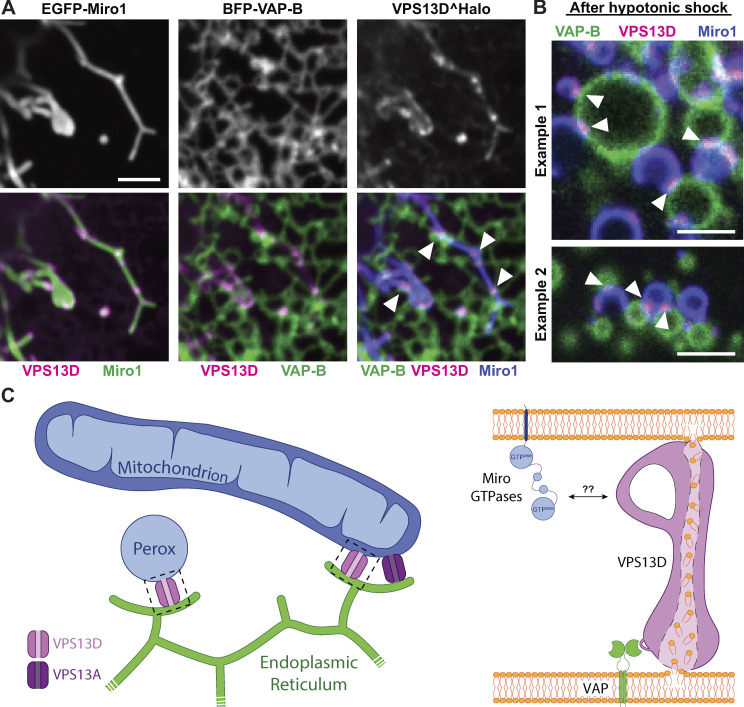
VPS13D can tether the ER to mitochondria in a VAP- and Miro-dependent way. (A) COS7 cells coexpressing EGFP-Miro1, BFP–VAP-B, and VPS13D^Halo. Top: Single fluorescence images. Bottom: Merge of the different fluorescence channels as indicated. White arrowheads in the triple merge show that VPS13D^Halo (magenta) is enriched at hot spots where the ER (green) and mitochondria (blue) intersect. (B) Vesiculated ER and mitochondria induced by hypotonic treatment of COS7 cells coexpressing VPS13D^Halo, GFP-Miro1, and BFP–VAP-B remain tethered to each other, and VPS13D^Halo concentrates at these sites (white arrowheads). See also Video 2. Scale bars, 3 µm. (C) Schematic cartoon summarizing key findings of this study. Left: VPS13D can bridge the ER and either mitochondria or peroxisomes. Right: The interaction of VPS13D with the ER is mediated by VAP, and its interaction with either mitochondria or peroxisomes is mediated by Miro. Based on the reported properties of VPS13 family proteins, it is proposed that VPS13D allows flux of lipids between the two tethered bilayers.

