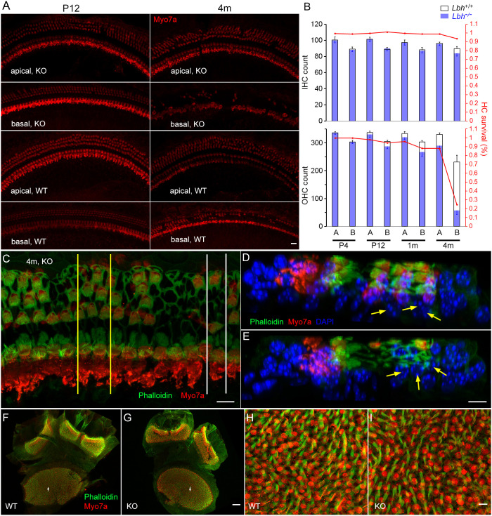
Fig. 3.
HC survival in the cochlear and vestibular sensory epithelia. (A) Representative confocal micrographs of HCs from an apical and a basal region in the cochleae of Lbh−/− and Lbh+/+ mice at P12 and 4 months. (B) IHC and OHC count from the two apical and basal areas (∼4.5 and 1.4 mm from the hook, each with 850 µm in length) of three Lbh−/− and three Lbh+/+ mice at P3, P12, 1 and 4 months. HC count from age marched Lbh+/+ mice was used as a reference and is presented as percentage of HC survival. A represents apical turn and B represents basal turn HCs in the plot. (C) Confocal image obtained from a 4-month-old Lbh−/− cochlea. Yellow lines and white lines mark the areas that were optical sectioned and are presented in D and E. (D,E) Optical sections of the two areas in C. Deiters’ cells’ nuclei are marked by yellow arrows. The nuclei of Deiters’ cells are still present at this stage despite the loss of OHCs. (F,G) Utricle macula and crista ampullaris of Lbh+/+ and Lbh−/− mice at 3 months. (H,I) Higher magnification images of areas marked by arrows in F and G. Scale bars: 10 µm (A,C-E,H,I); 50 µm (F,G).

