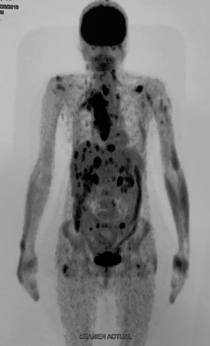
FIGURE 3.

The extent of metastatic disease following treatment was determined by 18F‐FDG uptake in the PET/CT scan conducted in August 2019 after 6 months of sorafenib treatment (reduced to 600 mg/day) and 5 months of lenvatinib treatment (20 mg/day). Multiple lymph node metastases were observed in the neck and mediastinum, with multiple secondary lesions in subcutaneous tissues and muscles, the liver, adrenal gland, and right pleura were identified (coronal view). 18 F‐FDG, 2‐deoxy‐2‐[fluorine‐18] fluoro‐D‐glucose; PET/CT, positron emission tomography/computed tomography
