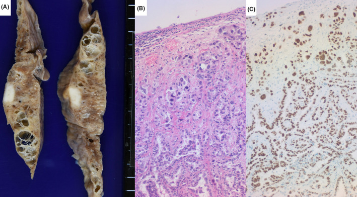
FIGURE 6.

Pulmonary adenocarcinoma in fibrotic lung parenchyma (a: gross appearance of right upper lobe, b: HE staining, c: thyroid transcription factor‐1 immunostaining). The 9‐mm white nodule is located just beneath the pleura. The surrounding lung tissue is fibrotic and honeycombed, focally resembling emphysema. Microscopically, well‐differentiated papillary adenocarcinoma shows neither necrosis nor lymphocytic infiltration. The nuclei of the cancer cells are diffusely immunoreactive for thyroid transcription factor‐1
