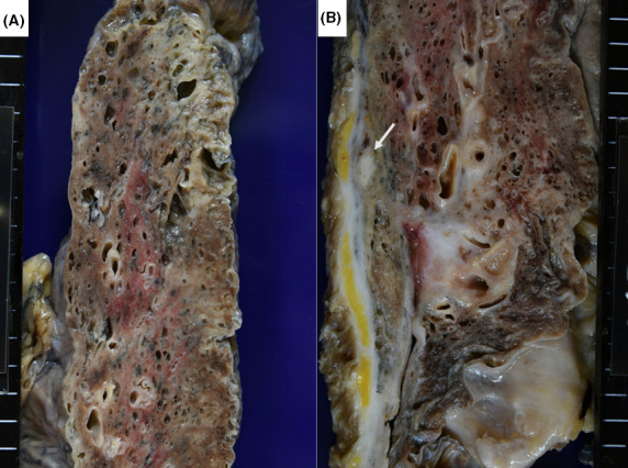
FIGURE 7.

Gross appearance of usual interstitial pneumonia (pulmonary fibrosis) with acute exacerbation (a: left upper lobe, b: right upper and middle lobes). Focal subpleural honeycombing and infiltrative change in the lung parenchyma are noted. The right pleura shows diffuse fibrous adhesion, and an encapsulated caseous focus is noted beneath the pleura of the collapsed middle lobe (arrow)
