
FIGURE 1.
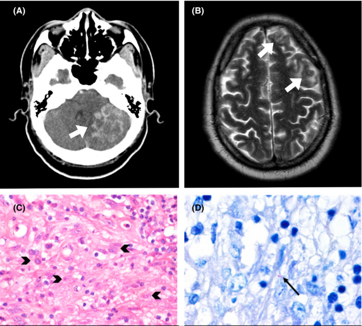
A, Axial brain computed tomography (CT) with contrast showing multiple ring‐enhancing lesions with perilesional edema and mass effect in the left cerebellar hemisphere (white arrow). B, Axial brain magnetic resonance imaging (MRI) with a heterogeneous lesion in the left frontal lobe with hypointensity on T2‐weighted image (white arrow). C, Brain histology with hematoxylin and eosin staining, 400× magnification, showed epithelioid histiocytes (black arrowheads). D‐ Ziehl‐Neelsen staining, 1000× magnification, revealed scattered bacilli (black arrow)