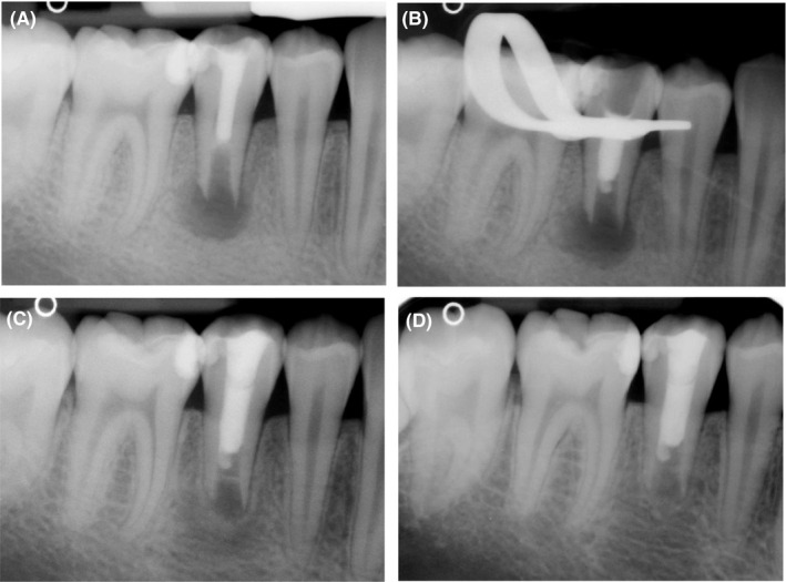
FIGURE 1.

Treatment of second mandibular right premolar. (A) Diagnostic radiograph that showed the presence of radiopaque intracanal and coronal filling associated with periapical radiolucent lesion. (B) Radiograph of tooth to check the placement of MTA barrier. (C) Six‐month radiographic follow‐up showing the complete resolution of periapical radiolucent lesion. (D) Twelve‐month radiographic follow‐up
