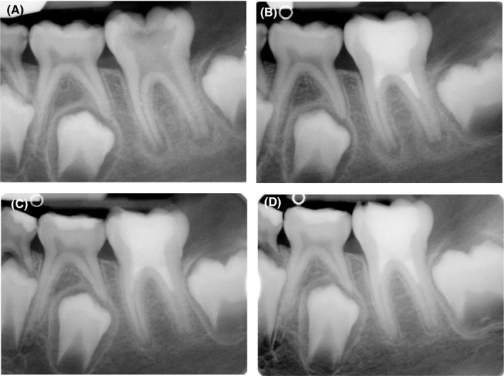
FIGURE 2.

Treatment of first left mandibular molar. (A) Preoperative radiograph of molar tooth. (B) Postoperative radiograph of first molar after treatment. (C) Six‐month radiographic follow‐up demonstrating the resolution of periapical lesion. (D) Twelve‐month follow‐up radiograph showing a partial apical closure of both roots
