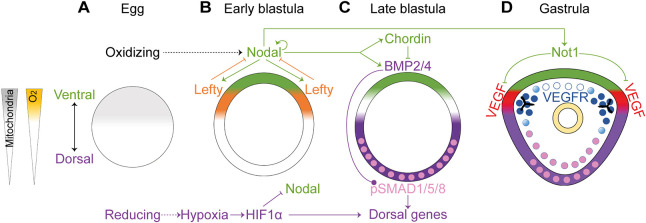
Fig. 1.
The regulation of DV axis formation downstream of redox and oxygen gradients in the sea urchin embryo. (A-D) Diagrams showing sea urchin DV and skeletal patterning in developing sea urchin embryos in normal conditions (based on Chang et al., 2017; Coffman et al., 2009; Coffman and Davidson, 2001; Coffman et al., 2004, 2014; Czihak, 1963; Duboc et al., 2004; Lapraz et al., 2009; Li et al., 2012). (A) The asymmetric distribution of mitochondria in the egg induces a redox gradient. (B) Regulatory interactions between nodal, lefty and HIF1α at the early blastula stage. (C) Nodal-mediated regulation of BMP signaling in the late blastula stage. (D) In the gastrula stage, Nodal activates the expression of Not1, which represses VEGF expression in the ventral ectoderm. Throughout the figure, the ventral side and Nodal expression domain are highlighted in green; the dorsal side and the domain of BMP activity are marked in purple. Nuclei that show pSMAD1/5/8 are highlighted in pink. VEGF expression is marked in red. VEGFR expression is marked in blue.

