Fig. 4.
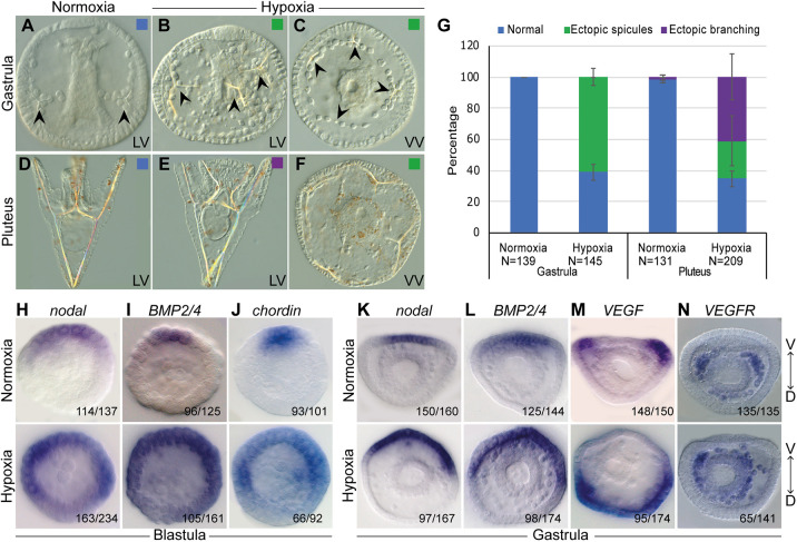
Growth in hypoxic conditions leads to skeletal defects and perturbs the expression of DV and skeletal patterning genes. (A-C) Representative images of embryos at gastrula stage. (A) Embryo grown in normoxic conditions shows normal development of two spicules (arrowheads). (B,C) Embryos grown in hypoxic conditions show ectopic spicules (arrowheads). (D-F) Representative images of embryos at pluteus stage. (D) Embryo grown in normoxic conditions shows a normal skeleton. (E) Embryo grown in hypoxic condition shows a normal DV axis and ectopic spicule branches. (F) Radialized embryo grown in hypoxic conditions that displays multiple ectopic spicules. LV, lateral view; VV, ventral view. (G) Quantification of skeletogenic phenotypes at gastrula stage and pluteus stage. Color code is indicated in the representative images. Error bars indicates s.d. of three independent biological replicates. (H-J) Spatial expression of nodal, BMP2/4 and chordin genes in normoxic (top) and hypoxic (bottom) embryos at blastula stage. (K-N) Spatial expression of nodal, BMP2, BMP4, VEGF and VEGFR genes in normoxic (top) and hypoxic embryos (bottom) at the gastrula stage. Embryos are presented in ventral view and the axis is labeled ventral (V) to dorsal (D). Throughout H-N, the numbers at the bottom right indicate the number of embryos that show this expression pattern out of all embryos scored, based on three independent biological replicates.

