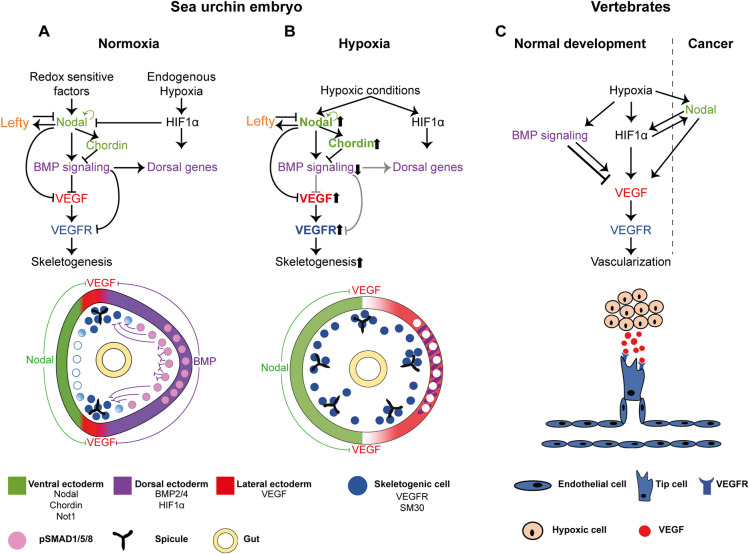
Fig. 7.
The interactions between the DV and skeletogenic GRNs, the response to early hypoxia and the similarities to the regulation of vertebrate vascularization. (A,B) Diagrams showing our proposed model for skeletal patterning in normal conditions (A) and hypoxic conditions (B). Color codes are indicated at the bottom of the figure. (A) The regulatory interactions between Nodal, BMP, HIF1α and VEGF signaling during normal development. BMP represses VEGF, VEGFR and SM30 expression in the dorsal side, and HIF1α does not regulate VEGF expression in the sea urchin embryo. (B) The modification of the regulatory states in hypoxic conditions applied at early development revealed in this work. Early hypoxia expands nodal expression and reduces BMP activity and the dorsal ectoderm. The reduction of BMP activity leads to an expansion of VEGF, VEGFR and SM30 expression in the dorsal side and to growth of ectopic skeletal centers. Upward arrows near a gene name indicate enhanced activity; downward arrows indicate reduced activity. Gray regulatory links indicate inactive connections under hypoxic conditions. (C) Diagram showing the relevant regulatory interactions during vertebrate vascularization in normal development and in cancer; see text for explanations.

