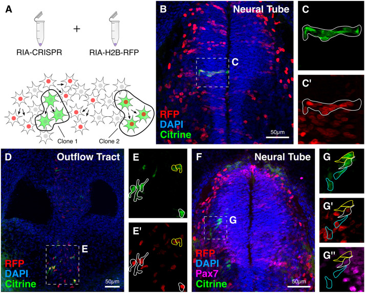
Fig. 9.
Proof-of-principle experiment to show application of RIA-CRISPR retroviruses for clonal analysis. (A) Experimental design for performing clonal analysis using RIA-CRISPR retroviruses. Clones were identified as single-infected cells with shared fluorescence intensity for Citrine, or double-infected cells with shared fluorescence intensity for Citrine and RFP. (B-E′) Examples of clonally related cells observed in the medial neural tube (B) and developing outflow tract (D). In the neural tube, clones (outlined) were horizontally distributed (C,C′), whereas in the outflow tract, clones were distributed evenly within the aorticopulmonary septum (E,E′). (F) In a representative embryo, double labeling with Citrine and RFP, together with intensity of Citrine and RFP and expression levels of Pax7, indicate clonal relationships. (G-G″) This allowed identification of three distinct clones: outlined in white – high Citrine (G), low RFP (G′), low Pax7 (G″); outlined in cyan – low Citrine (G), high RFP (G′), no Pax7 (G″); outlined in yellow – high Citrine (G), no RFP (G′), no Pax7 (G″).

