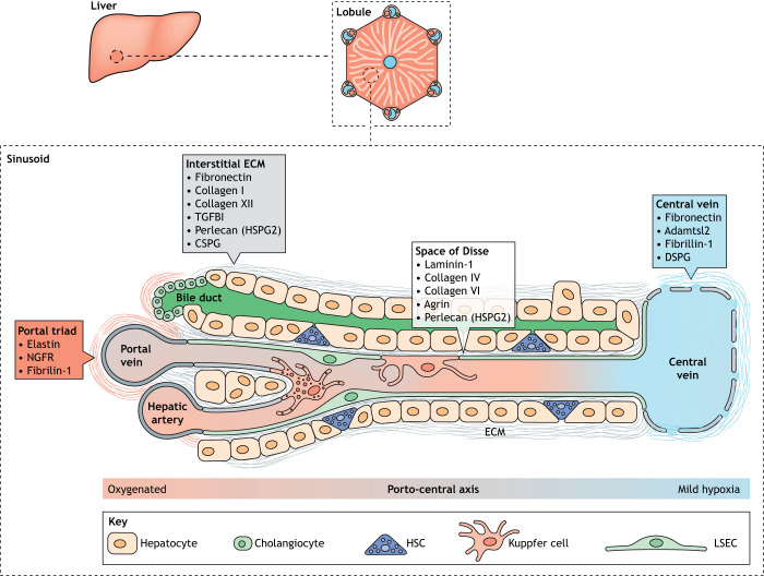
Fig. 1.
Liver structure, and cellular and common ECM proteins. Liver tissue is organised into discrete functional units called lobules that have a hexagonal arrangement of portal triads around a central vein, linked by sinusoids. LSECs line the sinusoid, whereas HSCs occupy the space of Disse that separates the sinusoid from hepatocytes. The different microstructures within the sinusoid have unique ECM compositions, shown in the boxes. A partial basement membrane lines the space of Disse, facilitating exchange of nutrients, proteins and xenobiotics. The interstitial matrix includes typical ECM components such as fibronectin and collagen I that support tissue structure and integrity. Differences also exist in ECM composition along the centroportal axis, although the functional relevance of this is unclear. CSPG, chondroitin sulfate proteoglycan 4; DSPG, dermatan sulfate proteoglycan; ECM, extracellular matrix; HSC, hepatic stellate cell; HSPG2, heparan sulfate proteoglycan core protein 2; LSEC, liver sinusoid endothelial cell; NGFR, tumour necrosis factor receptor superfamily member 16; TGFB1, transforming growth factor beta 1. The sinusoid and lobule structures are adapted from Frevert et al. (2005) and the Wikipedia Commons file 201904_hepatic_lobule.svg under the terms of the CC-BY 2.5 and 4.0 license, respectively.

