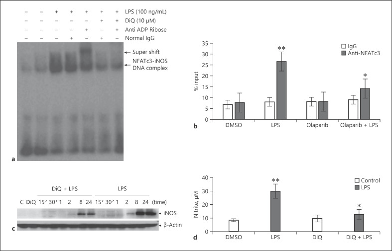
Fig. 2.
NFATc3 ribosylation increases binding of NFATc3 to iNOS gene promoter and gene expression. (a) Nuclear extracts from vehicle and LPS-stimulated BMDMs were incubated with a biotin-labeled NFAT consensus sequence from the iNOS promoter; separate binding reactions with DiQ, normal rabbit IgG, or anti-rabbit ADP-ribose antibody were analyzed by EMSA, and a representative blot is presented. (b) Macrophages were pretreated with vehicle (0.01% DMSO) or DiQ for 30 min and stimulated with LPS for 2 h. Treated cells were cross-linked with 1% formaldehyde, immunoprecipitated with anti-NAFTc3 or pre-immune serum, purified and used to amplify NFATc3 bound DNA on iNOS promoter by qPCR. (c) Expression levels of iNOS in vehicle or PARP-1 inhibitor pretreated cells that were stimulated with LPS for different time periods were determined by immunoblotting with anti-iNOS antibodies; (d) Release of NO into culture medium was measured by Greiss reagent. Statistical difference between different groups was calculated by 1-way ANOVA with Bonferroni correction. **p ≤ 0.01 LPS versus vehicle; *p ≤ 0.05 vehicle + LPS versus PARP-1 inhibitor + LPS. PARP-1, polyADP-ribose polymerase 1; NFATc3, nuclear factor of activated T-cell cytoplasmic member 3; BMDMs, bone marrow-derived macrophages; EMSA, electrophoretic mobility shift assay; DiQ, 1,5-isoquinolinediol; NFAT, nuclear factor of activated T cell.

