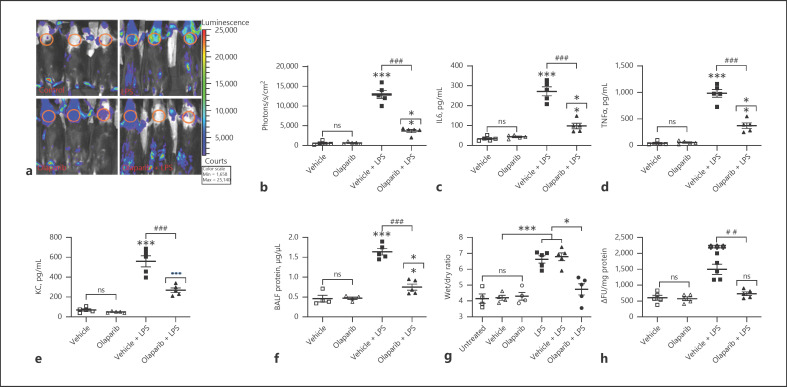
Fig. 5.
Prophylactic inhibition of NF-κB polyADP-ribosylation by olaparib inhibits LPS-induced ALI and pulmonary edema in NF-κB luciferase mice. NF-κB reporter mice were untreated, pretreated with 0.01% DMSO or olaparib (10 mg/kg) for 1 h, followed by intraperitoneal LPS (15 mg/kg) i.p. injection. At 16 h after LPS, ALI and pulmonary edema in the treated mice were measured by (a) bioluminescence imaging of the animals using an IVIS Lumina II imaging system. The circled chest area represents the region of interest for comparing the photon readings. b In vivo pulmonary inflammation was measured as photons emitted per second per cm2 mouse lung area. Similarly, parallel groups of NF-κB reporter mice were treated as described in Fig. 4, and BALF levels of IL-6 (c), TNFα (d), and KC (e), BALF protein (f), lung wet/dry ratios (g), and MPO levels in lung tissue were measured (h). Error bars represent mean ± SEM from 5 mice in each group. Statistical difference between different treatments was calculated by 1-way ANOVA with Bonferroni correction. ***p ≤ 0.001 vehicle + LPS or LPS alone versus vehicle or untreated control; ###p ≤ 0.001 vehicle + LPS versus olaparib + LPS; ##p ≤ 0.01 vehicle + LPS versus olaparib + LPS; **p ≤ 0.01 olaparib versus olaparib + LPS; •••p ≤ 0.001 olaparib versus olaparib + LPS; *p ≤ 0.05 untreated or vehicle + LPS versus olaparib + LPS (g). ALI, acute lung injury; BALF, bronchoalveolar fluid; MPO, myeloperoxidase.

