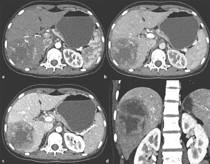
Fig. 1.
CASE 1: Triple-phase contrast-enhanced CT axial (a–c) and coronal (d) images: in arterial phase, mass in right lobe shows peripheral arterial enhancement (a), portal venous (b), hepatic venous phases − progressive enhancement is seen in the periphery of the tumor with cystic nonenhancing areas in the center of the tumor (c, d). The rest of the liver shows normal enhancement.

