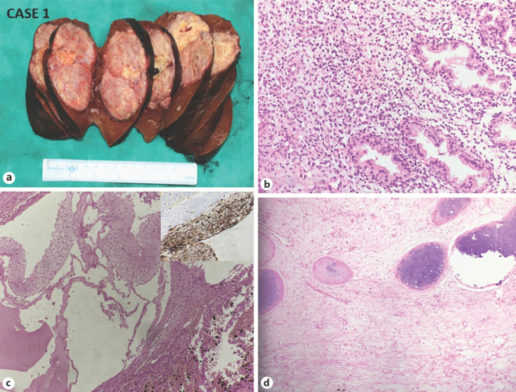
Fig. 2.
CASE 1: Mixed HB with the teratoid feature. a Gross: well-defined tumor nodule, abutting the capsular surface (H and E, ×200). b Microphotograph shows an epithelial component of HB. c Microphotograph shows Glial component and melanin pigment (H and E, ×200) (right upper inset showing GFAP immunopositivity). d Microphotograph shows osteoid and cartilage formation H and E, ×200). HB, hepatoblastoma.

