Fig. 4.
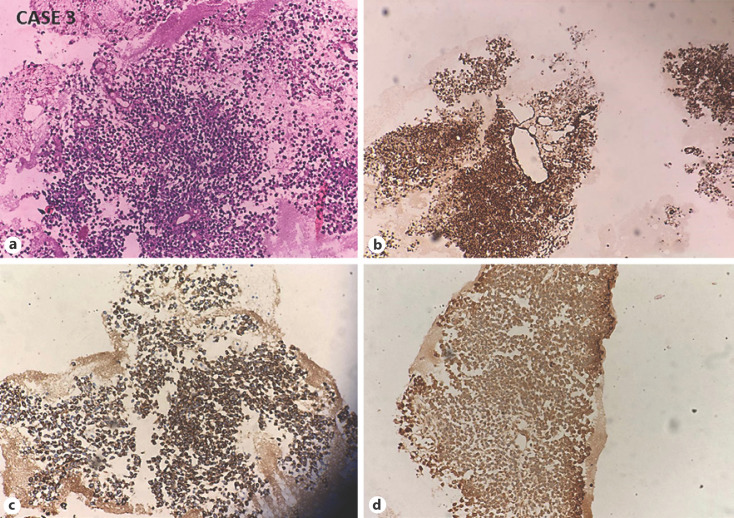
CASE 3: Small-cell undifferentiated HB. a Microphotograph shows sheets and perivascular arrangement of dyscohesive small blue cells with scant cytoplasm (H and E, ×200). Tumor cells are immunoreactive for cytokeratin (b), vimentin (c), and focally for alpha-fetoprotein (d). HB, hepatoblastoma.

