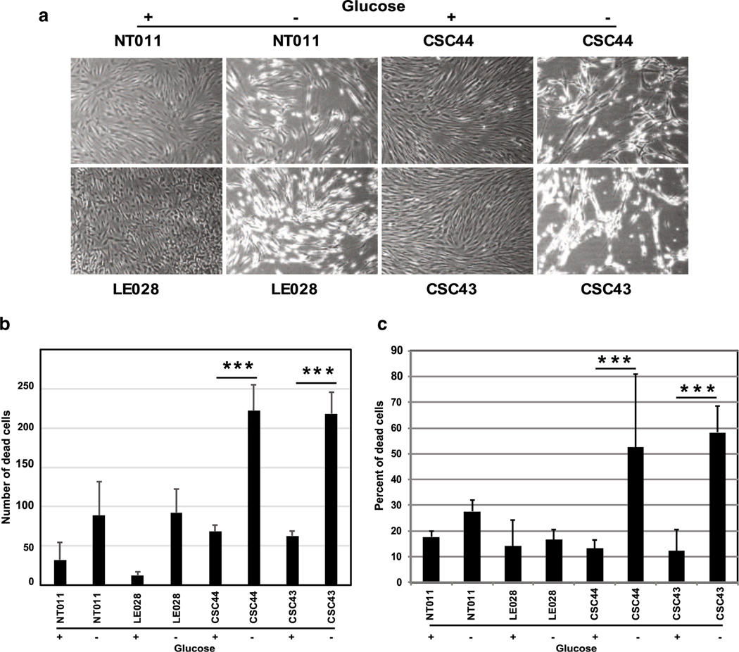
Fig. 5.
Effect of glucose deprivation on cell death. Dermal fibroblasts from normal donors (NT011 and LE028) and patients (CSC43 and 44) were grown in media containing normal media (indicated by +) or media lacking glucose (indicated by −) for 6 days. The cells were photographed under phase contrast (a) or quantitated under fluorescence microscope after staining with DAPI (b). Cells were also stained with Annexin V and PI using an apoptosis detection reagents and analyzed by FACS (c). The experiments were repeated 3 times and the average number and percentage of cell death with standard deviation was plotted. The P values were determined by comparing the cell death in the presence and absence of glucose in growth medium:***P < 0.001

