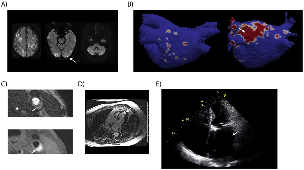
Figure 3. Imaging Findings Associated with Potential Occult Sources of Currently Unexplained Ischemic Stroke.

(A) Diffusion-weighted magnetic resonance imaging demonstrating acute infarction in all three major arterial territories of the brain, a finding suspicious for cancer-related hypercoagulability. (B) Cardiac magnetic resonance imaging demonstrating significant areas of atrial fibrosis (image on right) compared to an atrium with minimal fibrosis (image on left).73 (C) Hemorrhage within nonstenosing atherosclerotic plaque demonstrated on 3-dimensional time-of-flight magnetic resonance angiography (upper image) and T1 CUBE FS sequences (lower image). (D) Cardiac magnetic resonance imaging demonstrating late gadolinium enhancement consistent with MI in an ESUS patient with no clinical history of MI. (E) Echocardiography demonstrating a severely dilated left atrium in an ESUS patient without known atrial fibrillation.
