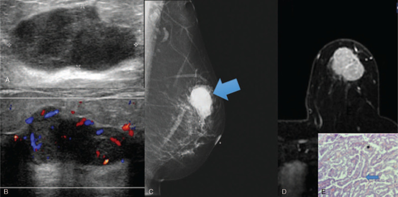
Figure 1.

Invasive papillary carcinoma solid mass sonographic appearance. 50 year old lady presented with a mass in the left breast. Figure 1A showed a lobulated hypoechoic mass, with increased vascularity on Doppler images (Fig. 1B). Mammogram (Fig. 1C) demonstrates a high density mass in the left upper quadrant. Figure 1D is of subtracted MRI image post gadolinium, with a large heterogeneously enhancing mass which shows washout on the delayed phase (Type 3 curve). Histopathological examination (Figure 1E) demonstrates numerous papillary structures (blue arrow). (Magnification 40x).
