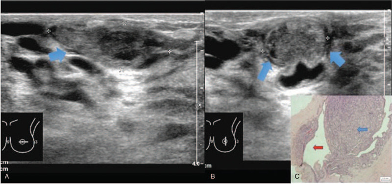
Figure 2.

Intraductal papilloma solid mass with anechoic rim sonographic appearance. 30 year old lady presented with left nipple discharge. Figure 2A and 2B showed a solid mass with anechoic rim (arrows) on ultrasound images. Doppler image (Fig. 2C) of the mass revealed peripheral but no internal vascularity. Histopathological examination (Fig. 2D) demonstrates intraductal papilloma (blue arrow) within a duct (red arrow). (Magnification 20x).
