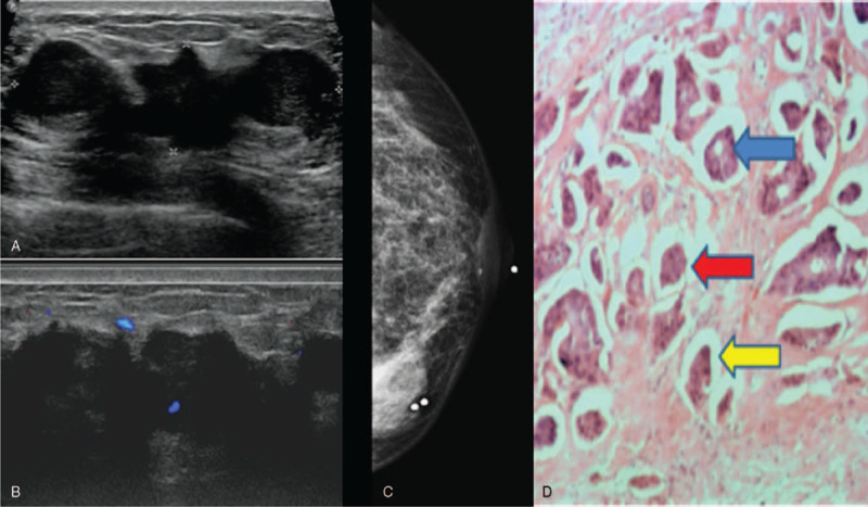
Figure 5.

Invasive micropapillary carcinoma solid mass appearance. 49 year old lady presented with left sided nipple discharge. Figure 5A showed a large solid hypoechoic mass which was lobulated with irregular margins. Doppler images demonstrated increased vascularity (Fig. 5B). Mammogram (Fig. 5C) showed a high density mass with irregular margins in the left lower inner quadrant. Histopathological examination (Fig. 5D) demonstrates invasive tumor cells in vague glands [blue arrow] and nests [red arrows], most of which appear within clear spaces [yellow arrow]. (Magnification 40x).
