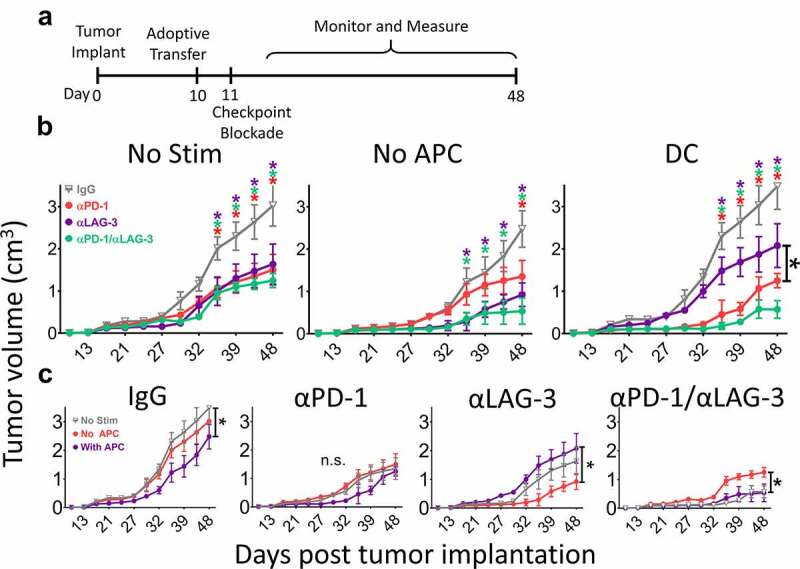
Figure 2.

Blockade of PD-1 or LAG-3 improves anti-tumor activity of activated CD8 + T-cells. As shown in panel A, B6 mice were inoculated with 1 × 10⁶ PD-L1-expressing E.G7-OVA cells. After ten days, 1 × 10⁶ OT-1 T cells, stimulated with or without peptide and with or without DC as in Figure 1, were adoptively transferred into the tumor-bearing mice. The following day, mice were treated with IgG isotype control (gray), PD-1 blocking (red), LAG-3 blocking (purple), or a combination of both PD-1 and LAG-3 blocking antibodies (green). Tumor growth was measured as indicated on the X axes. Shown in panel B are the growth curves for mice that received T cells which had not been incubated with DC and a nonspecific peptide (No Stim), T without DC cells stimulated with SIINFEKL peptide alone (No APC), or T cells stimulated with peptide in the presence of DC (DC). Panel C shows the same data grouped by checkpoint blockade treatment rather than T-cell stimulation conditions. Measurements for individual mice are shown in Supplemental Figure S5. Asterisks indicate p < .05 as assessed by 2-way ANOVA with Bonferroni’s multiple comparisons test. Results are from one experiment with N = 6 mice per group
