
Figure 5.
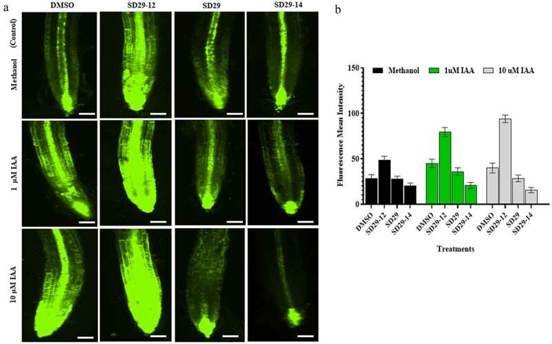
The RACK1A modulators mediate auxin signal in DR5rev::GFP. Roots of seven-day-old, containing DR5rev::GFP construct, were treated as follows: (a) Control set including methanol (m) plus DMSO (d), SD29-12, SD29 and SD29-14, respectively. Treatment sets supplemented with 1 μM or 10 μM IAA plus D, SD29-12, SD29 and SD29-14, respectively. (b) Quantification of the lateral roots’ numbers after each treatment. The fluorescence signal was detected using Eclipse Ti 2000 laser-scanning confocal microscope (Nikon CSU series Spinning Disk confocal microscope). with FITC filter set. The roots were observed under a Nikon confocal microscope (20× magnification lens). The argon (488 nm) laser with appropriate emission filters was used for the visualization of FITC. The expression of GFP was quantified using ImageJ software. The black pars were controls (DMSO, SD29-12, SD29 and SD29-14) with methanol, the green pars were treatments (DMSO, SD29-12, SD29 and SD29-14) with 1 μM IAA, the gray pars were treatments (DMSO, SD29-12, SD29 and SD29-14) with 10 μM IAA. Error bars indicate standard deviation from the mean n = 3 independent experiments