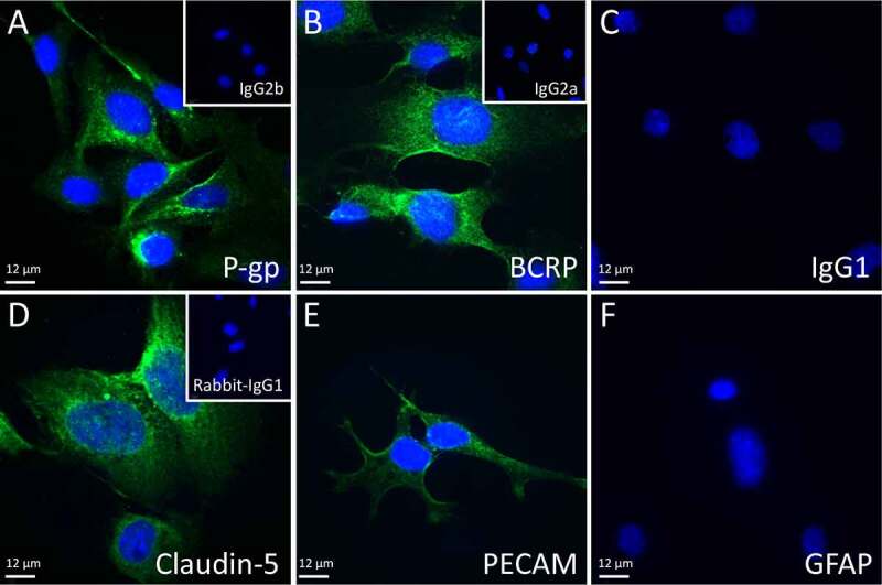
Figure 1.

Representative fluorescent immunohistochemical images used for hCMEC/D3 characterization. Blue indicates DAPI staining, green represents specific antibody staining: A) P-gp, B) BCRP, C) IgG1, D) Claudin-5, E) PECAM, and F) GFAP. Insets show IgG2b (a), IgG2a (b) and rabbit IgG (d), 40X objective. Scale bars = 12 µm
