
Figure 3.
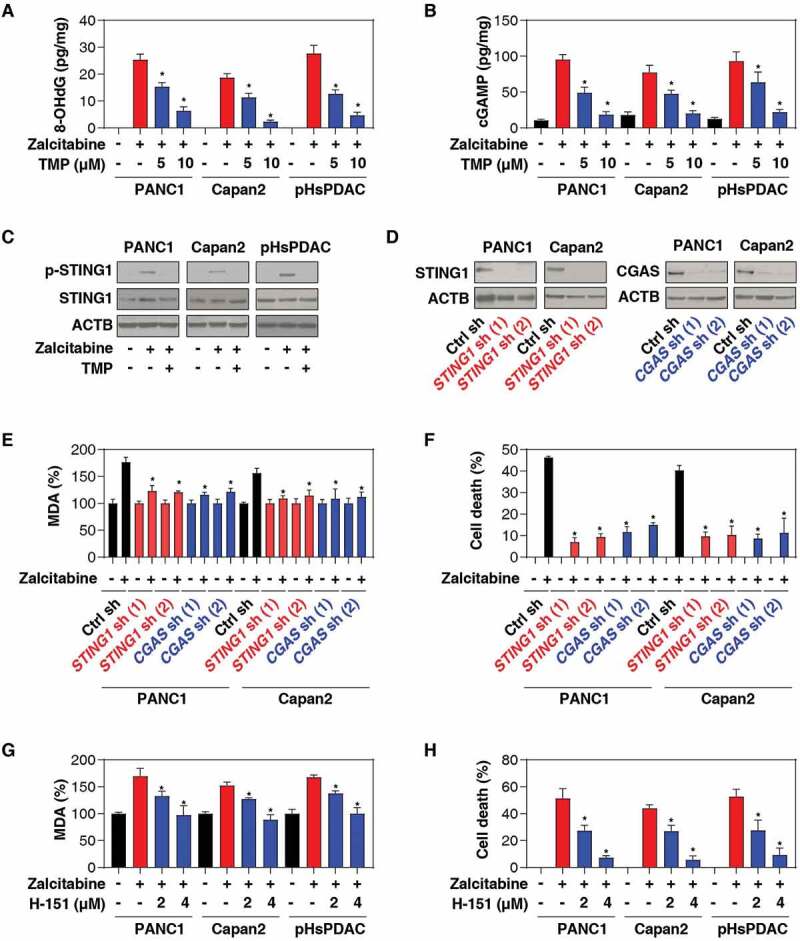
The CGAS-STING1 pathway promotes zalcitabine-induced ferroptosis. (A-C) The indicated human PDAC cells were treated with zalcitabine (20 µM) in the absence or presence of TMP (5 or 10 µM) for 72 h. The intracellular levels of 8-OHdG (A), cytosolic mtDNA (B), and cGAMP (C) were assayed with ELISA kits (n = 3, *P < 0.05 versus zalcitabine-alone group). (D) Western blot analysis of protein expression in human PDAC cells following treatment with zalcitabine (20 µM) in the absence or presence of TMP (10 µM) for 72 h. (E) Western blot analysis of protein expression in the indicated STING1- and CGAS-knockdown PDAC. (F, G) Analysis of levels of MDA (F) and cell death (G) in the indicated STING1- and CGAS-knockdown PDAC cells following treatment with zalcitabine (20 µM) for 72 h (n = 3, *P < 0.05 versus control shRNA group). (H-I) Analysis of levels of MDA (H) and cell death (I) in the indicated PDAC cells following treatment with zalcitabine (20 µM) in the absence or presence of the STING1 inhibitor H-151 (2 or 4 µM) for 72 h (n = 3, *P < 0.05 versus zalcitabine-alone group)