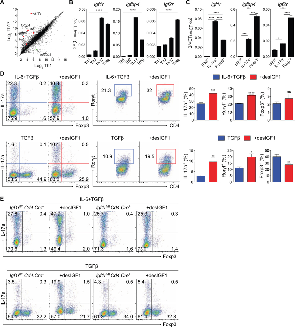
Figure 1. Signaling through IGF1R reciprocally regulates Th17 and Treg cell differentiation in-vitro.
(A) Scatterplot of gene expression microarray analysis of RNA from Th1 and Th17 cell conditions. Each data point represents the average of all probes above background for a given gene. (B-C) Sorted naive CD4+ T cells were stimulated in-vitro for 5 days in Th1, Th2, Th17 or Treg cell conditions. (B) qPCR analysis of Igf1R, Igfbp4 and Igf2r mRNA isolated from sorted total WT cells. (C) qPCR analysis of Igf1R, Igfbp4 and Igf2r mRNA (bottom) isolated from sorted reporter-positive IFNγ.Thy1.1-IL-17a.hCD2-Foxp3.GFP cells. (top). Experiments performed 3 times. Data from one representative experiment shown. (D-E) Sorted naive CD4+ T cells from WT (D) or IFGR1-deficient (E) mice were stimulated in-vitro for 5 days in Th17 or Treg cell conditions with or without desIGF1 and subjected to flow cytometric analysis for IL-17a, Foxp3, and RORγt. (D) Dot plots (left panels) and bar plots (right panels) are concatenated from three replicates in a single experiment. Data representative of 3–5 separate experiments. (E) Flow cytometry plots of IL-17a and Foxp3 expression from Igf1rfl/fl Cd4.Cre– and Cd4.Cre+ cells, depicting cell-number controlled concatenated data of three replicates from one of three similar experiment. Student’s t-tests were used to assess significance. See also Supplementary Figs. S1 and S2.

