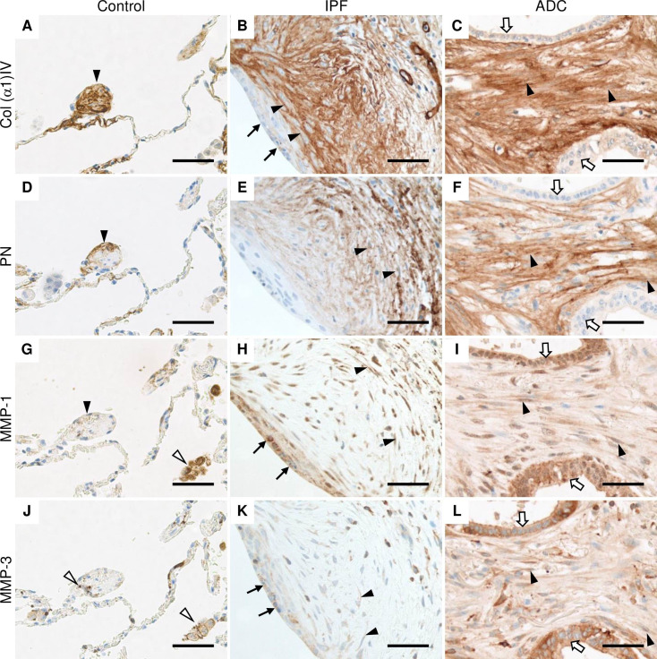
Fig 2. Immunohistochemical localization of Col α1(IV), PN, MMP-1 and MMP-3 in normal control lung, IPF and ADC.
Sections were obtained from patients with lung adenocarcinoma (ADC) from both the tumor area and histologically normal lung as well as from patients with idiopathic pulmonary fibrosis (IPF). (A, B and C) Stromal cells of control lung, IPF and ADC were strongly positive for collagen (Col) α1(IV) chain (black arrow heads), while hyperplastic alveolar epithelial cells lining fibroblast focus (black arrows) and cancer cells (white arrows) were weakly positive. (D, E, and F) Stromal cells of control lung, IPF and ADC were positive for periostin (PN) (black arrow heads). (G) Alveolar macrophages (white arrow heads) were strongly positive for matrix metalloproteinase (MMP) -1, while cells with a widened alveolar tip of normal lung were weakly positive (black arrowhead). (H) Stromal cells (black arrow heads) and epithelial cells lining fibroblast foci (black arrows) in IPF were positive for MMP-1. (I) Cancer cells (white arrows) and stromal cells (black arrow heads) of ADC were positive for MMP-1. (J) Alveolar macrophages and monocyte lineage cells (white arrow heads) were positive for MMP-3 in normal lung. (K) Hyperplastic alveolar epithelial cells (black arrows) and some stromal cells (black arrow heads) were positive for MMP-3 in IPF. (L) Stromal cells (black arrow heads) as well as cancer cells (white arrows) were positive for MMP-3. Scale bar 50 μm.

