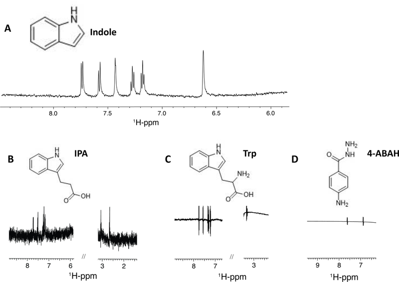
Figure 4. Indole metabolites exhibit ligand binding to myeloperoxidase by NMR Saturation Transfer Difference (STD).
1D 1H-STD NMR spectra of 9 μM rhMPO in association with 1 mM aqueous solutions of the ligands (A) indole, (B) indole-3-propionic acid, (C) tryptophan, (D) 4-ABAH. The corresponding ligand structure is shown on top of each spectrum.

