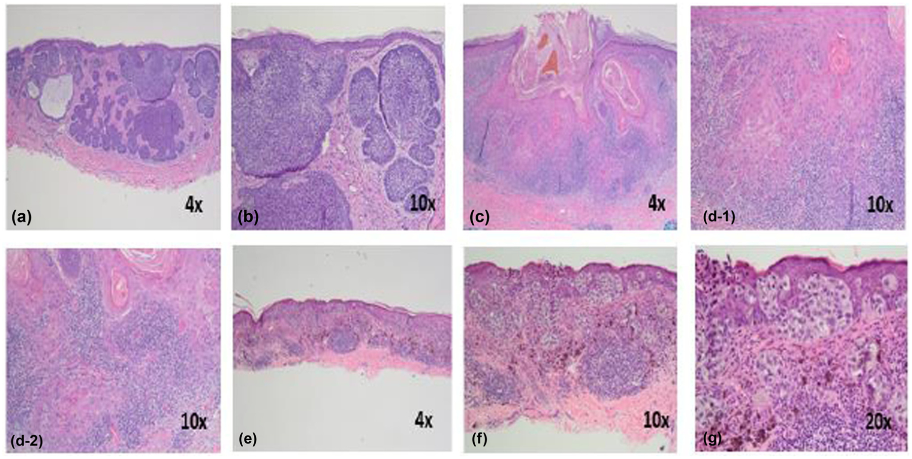
Figure 1:

The characteristic histology of different forms of skin cancer: basal cell carcinoma: (a) nodular masses of basaloid cells extend into the dermis with peritumoral stroma (4×); (b) closer magnification highlights the peripheral palisading and tumor retraction artifact (10×); squamous cell carcinoma: (c) scanning magnification highlights a crater-like tumor with irregular masses of atypical keratinocytes invading the dermis. A pronounced surrounding inflammatory infiltrate is seen (4×); (d-1) and (d-2) invading tumor masses with atypical keratinocytes, dyskeratotic cells, and keratin pearls (10×); melanoma: (e) scanning magnification reveals a broad, atypical melanocytic proliferation which is asymmetric and poorly circumscribed (4×); (f) the epidermal proliferation is characterized by variably sized nests at and above the dermoepidermal junction. A robust lymphocytic inflammatory response is present (10×); (g) closer magnification again reveals nests at and above the dermoepidermal junction. The lesional cells are atypical, and single melanocytes are noted at all levels of the epidermis in a pagetoid arrangement (20×) (photographs were provided by Dr. Allison Cruse, Department of Dermatology, University of Mississippi Medical Center, Jackson, Mississippi, USA).
