
Figure 1: Distribution of Selected Coronary Vascular Function Testing in 263 Women with Evidence of Ischemia and no Obstructive Coronary Artery Disease.
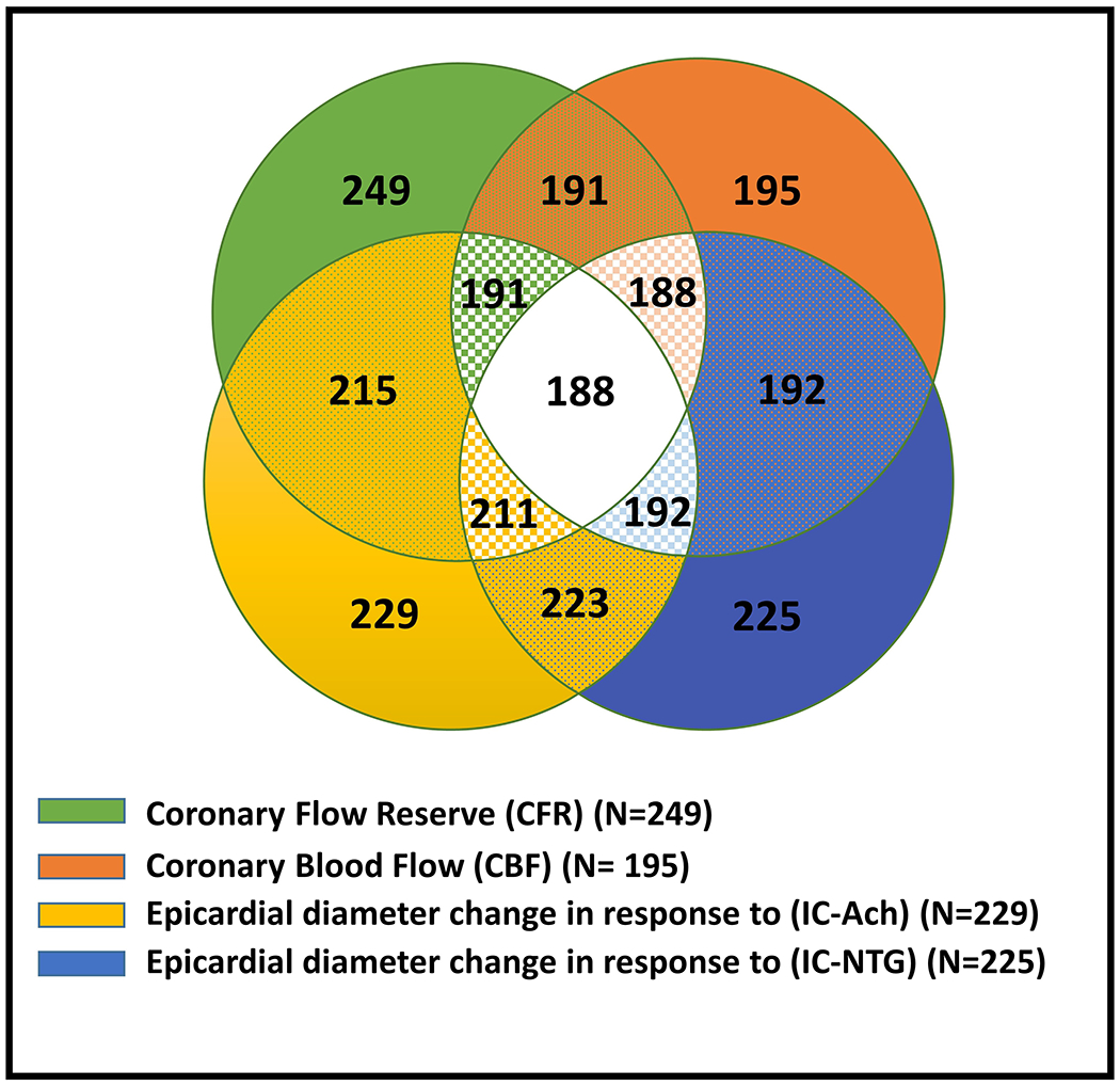
Most women underwent evaluation of >1 coronary vascular function pathway. Green represents women who underwent evaluation of coronary microvascular dilation using coronary flow reserve (CFR); orange represents women who underwent evaluation of coronary microvascular constriction using coronary blood flow (CBF); yellow represents women who underwent evaluation of coronary epicardial constriction using change in coronary artery diameter in response to intracoronary acetylcholine (IC-Ach); blue represents women who underwent evaluation of coronary epicardial dilation using change in in response to intracoronary nitroglycerin (IC-NTG).