
Fig. 1.
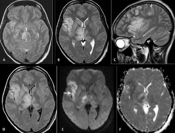
Cranial magnetic resonance imaging was done on day 9 on 28 female patients with acute encephalitic symptoms. Axial T2WI ( A and B ) and sagittal T2WI ( C ) images showing asymmetrical hyperintensities in bilateral substantia nigra and thalami with the affection of the right insular cortex. Axial fluid-attenuated inversion recovery image ( D ) also showing the abnormalities. Axial diffusion-weighted imaging ( E ) and apparent diffusion coefficient ( ADC ) map ( F ) images showing diffusion restrictions in the affected regions with low ADC value (arrow).