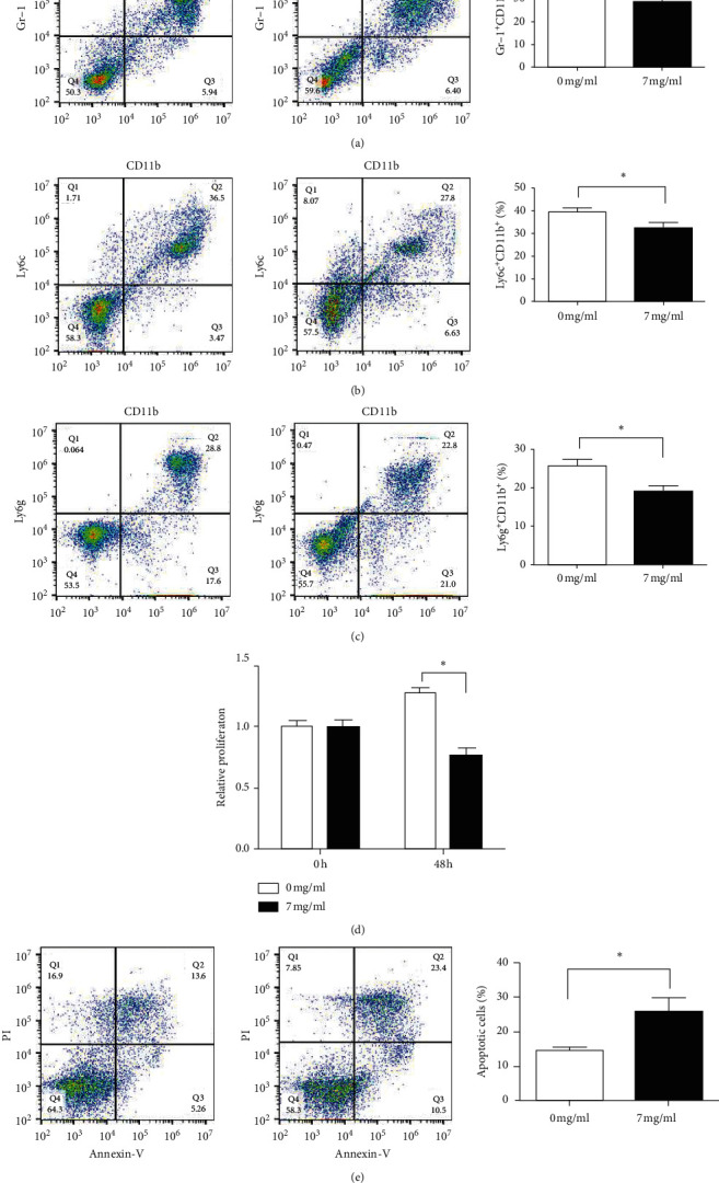
Figure 4.

Effects of the YPF formula on proliferation and apoptosis of MDSCs and its subsets. (a) Proportion of MDSCs (Gr-1+ CD11b+). (b) The proportion of monocytic MDSCs (Gr-1+CD11b+Ly6G−Ly6Chi). (c) The proportion of granulocytic MDSCs (Gr-1+CD11b+Ly6G+Ly6Clo). (d) The proliferation of MDSCs was detected by the CCK-8 assay. (e) Apoptotic cells were routinely detected by the Annexin V/PI staining method. The proportion of apoptotic cells is shown in the lower right quadrant, and the cells shown in the upper right quadrant are necrotic cells or cells with secondary necrosis after apoptosis. The concentration of the YPF formula was 0 mg/mL for the control group and 7 mg/mL for the YPF group. ∗P < 0.05 vs. control.
