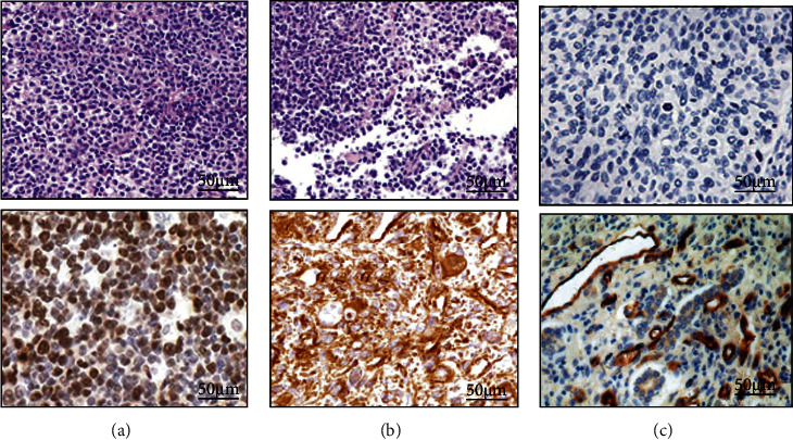
Figure 3.

The immunohistochemical results of PD-1 and PD-L1 (SP × 200). (a–c) were tumor cells, cancer mesenchymal cells, and tumor-infiltrating lymphocytes, respectively. A1 was PD-L1 (negative); A2 was PD-L1 (positive); B1 was PD-L1 (negative); B2 was PD-L1 (positive); C1 was PD-1 (negative); C2 was PD-1 (positive).
