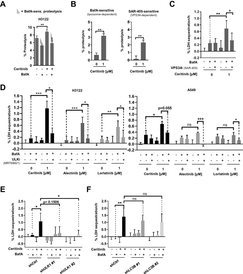
Figure 2.
Ceritinib triggers ULK1- and VPS34-dependent autophagy (A) Bar plot represents percent proteolysis of H3122 cells after treatment with DMSO or Ceritinib for 24 h in the presence or absence of BafA during the last 5 h (n = 3). (B) Quantification of BafA-sensitive and SAR-405-sensitive proteolysis in H3122 cells after treatment with 1 µM Ceritinib for 24 h (n = 3). Percent proteolysis was determined and calculated as described in the methods section. (C) H3122 cells were treated with DMSO or 1 µM Ceritinib (24 h) ± BafA and ± SAR405 (during the last 3 h) before LDH sequestration was determined (n = 3). (D) H3122 and A549 cells were treated with DMSO or 1 µM Ceritinib/Alectinib/Lorlatinib (24 h) ± BafA and ± MRT68921 (during the last 3 h) before LDH sequestration was determined (n = 3). (E) H3122 control (shCtrl) and two ULK1 knock down (shULK1#1 and shULK1#2) cell lines were subjected to LDH sequestration assay after 18 h of Ceritinib treatment. BafA was added during the last 3 h (n = 3). (F) Experiment as in E, but with H3122 shCtrl, shLC3B#1 and shLC3B#2 cells (n = 3). Mann–Whitney U was applied to compare two groups; *p < 0.05, **p < 0.01, ***p < 0.001.

