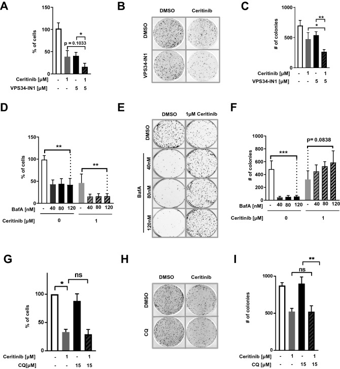
Figure 3.
Blocking autophagy via VPS34 inhibition sensitizes cells to Ceritinib (A) H3122 cells were treated with DMSO or 1 µM Ceritinib in the presence or absence of VPS34-IN1 for 2 days before living cells were counted and re-seeded for clonogenic assays. Bar plot represents percent of living cells compared to control treated cells (n = 4). (B) Clonogenic assay of experiments as described in A. After 2 days of treatment, 5 × 10³ living cells were re-seeded and kept for 10 days without treatment. Thereafter, colonies were stained with crystal violet and counted. (C) Quantification of the clonogenic assays described in A (n = 4). (D) H3122 cells were treated with DMSO or 1 µM Ceritinib in the presence or absence of increasing concentrations of BafA (40, 80 and 120 nM) for 2 days before living cells were counted and quantified from 5 independent experiments as in A. (E) Clonogenic assay of cells as described in D. Living cells were re-seeded at low density (5 × 10³ cells per well in a 6-well plate) and cultured in the absence of drugs for 10 days. Colonies were counterstained using crystal violet and counted. (F) Quantification of colonies shown in E (n = 5). (G) H3122 cells were treated with DMSO or 1 µM Ceritinib in the presence or absence of 15 µM chloroquine and asssessed as in A (n = 3). (H) Clonogenic assay of chloroquine (CQ) treated cells as described in G and assessed as in B. (I) Quantification of colonies shown in H (n = 3).

