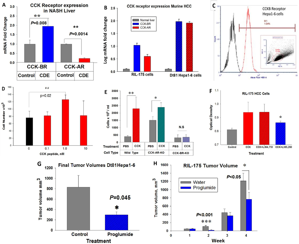
Figure 1.
Expression of CCK receptors in the murine liver and HCC cells and response to CCK. A, CCK-BRs are significantly increase in the NASH livers of mice on the CDE diet compared to livers of control mice (P=0.008). CCK-ARs are downregulated compared to control livers in mice fed a high saturated fat CDE diet (P=0.0014). B, Compared to normal mouse liver, CCK-BR and CC-ARs are significantly increased in mouse RIL-175 and Dt81Hepa1-6 HCC cells. C, CCK-BR immunofluorescence is detected by flow cytometry in Dt81Hepa1-6 HCC cells. †reproduced with permission from Dig Dis Sci (21). D, CCK peptide stimulates growth of Dt81Hepa1-6 HCC murine cells in cell culture with the greatest effect with 1nM, a concentration equal to the physiologic binding affinity at the CCK-BR. E, Means ± SD of Dt81Hepa1-6 wild-type, CCK-AR-KO or CCK-BR-KO cells after four days of exposure to PBS or CCK (1 nM). CCK peptide stimulated growth of wild-type cells and CCK-AR-KO cells but not CCK-BR-KO cells. F, CCK (1 nM) stimulated growth of RIL-175 murine HCC cells compared to controls. The CCK-increased growth was blocked only by the CCK-BR antagonist, L365,260, and not the CCK-AR antagonist, L364,718. G, In vivo, Dt81Hepa1-6 tumor size was significantly less in mice treated with proglumide compared to controls. H, In vivo, weekly tumor volumes of RIL-175 murine HCC tumors were significantly less in mice that received drinking water supplemented with proglumide. Significantly different than controls: *P<0.05, **P<0.01, ***P<0.001.

