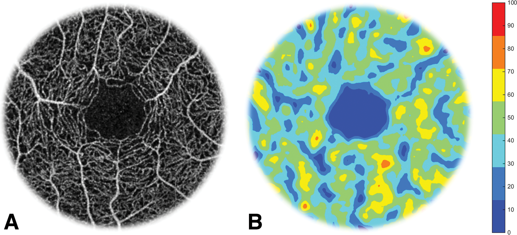
Fig. 3. Foveal Avascular Zone (FAZ) and perfusion map.

The enface angiography vessel image was obtained using optical coherence tomography (OCT)angiography (Avanti OptoVue; OptoVue). The FAZ is located in the center (the dark area) of the macula in the enface angiography of the retina (A). To visualize capillary perfusion, a Gaussian density filter (4 × 4 pixels) was used to process the skeletonized vascular images and the capillary density was color coded to create a capillary perfusion map (B), which shows the non-perfusion in the center of the fovea. The bar denotes to percent regional perfusion rate.
