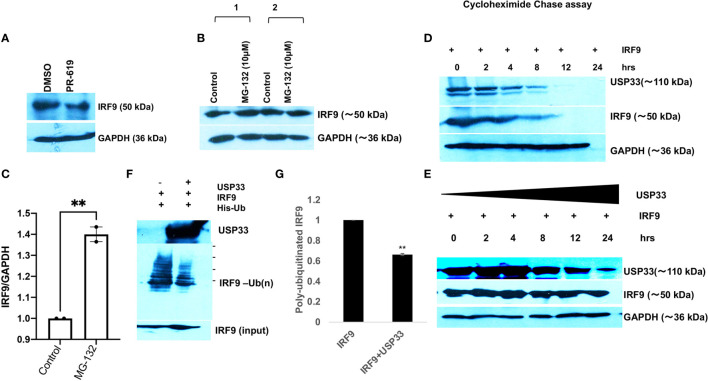
Figure 5.
USP33 regulates the IRF9 turnover via its deubiquitylation in microglia. (A) Immunoblot image showing the impact of deubiquitinase inhibitor PR-619 on the levels of IRF9 in microglial cells. PR-619 were treated at 10 µM final concentration for 8 hours and cells were harvested and followed by western blot analysis. (B) Immunoblot images showing the impact of proteasome inhibitor MG-132 on the elevated stability of IRF9 in microglial cells. (C) Graph bar displaying the average increase in IRF9 levels upon MG-132 treated human microglial cells. (D) Immunoblot analysis of half-life of IRF9 protein measured via cycloheximide chase assay done in HEK-293T cells. (E) Immunoblot analysis of impact of USP33 on turnover of IRF9; measured via cycloheximide chase assay performed in HEK-293T cells. Cycloheximide chase assay have been repeated two times and representative results are displayed here. (F) Immunoblot image showing the impact of USP33 on polyubiquitinated levels of IRF9. 5µg of pLV-IRF9 plasmids and 5µg of 6X-His-ubiquitin plasmids were transfected along with 5µg of Flag-HA-USP33 in co-transfection experiments (in 60mm dish format) to check the impact of USP33 on ubiquitination levels of IRF9. After 24 hours, cells were treated with MG132 for 8 hours, followed by in vivo ubiquitination assay as described in detail in Material and Methods section. (G) The graph bars showing the quantified levels of poly-ubiquitinated IRF9. The ubiquitination assay was performed three times independently and average values are shown as graph bars. Data are displayed as mean and mean ± S.E.M. student’s t-test have been utilized to obtain the statistical significance, indicated as ** for p values <0.005.

