Figure 1.
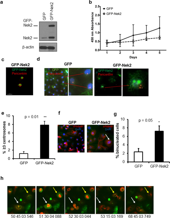
Nek2 overexpression causes centrosome amplification and binucleation. (a) Representative immunoblot of MCF10A cells expressing either GFP or GFP-Nek2 indicating overexpression of endogenous Nek2 and GFP-Nek2 detected with Nek2 antibodies. (b) CCK8 cell growth/viability assay comparing MCF10A expressing GFP or GFP-Nek2. (c) High-resolution microscopy of a centrosome pair indicating localization of pericentrin (red) and GFP-Nek2 (green). (d) Immunofluorescent staining of pericentrin (red) in GFP and GFP-Nek2-expressing cells (green) counterstained with DAPI (blue). The right inset (GFP-Nek2) indicates a cell with 3 centrosomes. (e) Quantifications of fluorescent microscopy for centrosome amplification by pericentrin staining. N = 3, bars = mean ± SD, *p < 0.05. (f) Immunofluorescent staining of α-tubulin (red) in GFP and GFP-Nek2-expressing cells counterstained with DAPI (blue). The arrow indicates a binucleated cell. (g) Quantifications of fluorescent microscopy for binucleation by α-tubulin staining. N = 3, bars = mean ± SD, *p < 0.05. (h) Representative screenshots of MCF10A/GFP-Nek2 cells subjected to live imaging for 72 h. Centrosome-localized GFP-Nek2 is visible in green and the microtubules are marked by RFP-α-tubulin. Arrows indicate tripolar cell division at different time points.

