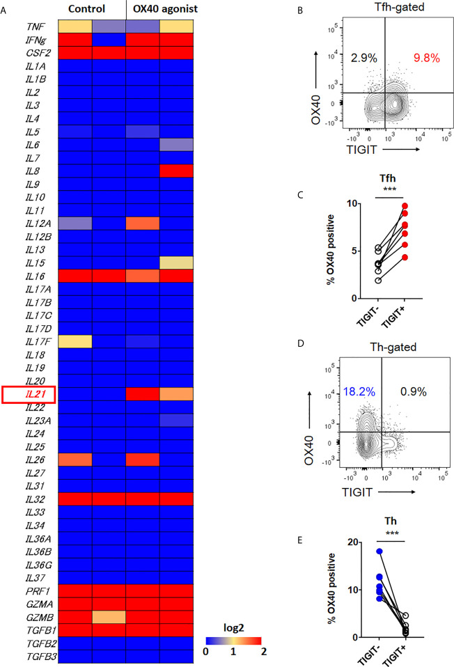
Figure 6.
OX40 signal is associated with IL-21 expression. (A) CD4+ T cells were stimulated in vitro in the presence of αCD3/CD28 activation beads with or without agonistic OX40 antibody for 3 to 5 days. Transcriptome analysis for 49 cytokines was obtained by RNA-sequencing, and the gene expression levels are shown as a heatmap. (B, D) OX40 expression level was examined in CXCR5+Tfh and CXCR5-Th cells by flow cytometry. (B, D), representative image. (C, E), multiple comparison results (n=7). (C, E) paired t-test. ***p<0.001.

