
Figure 6.
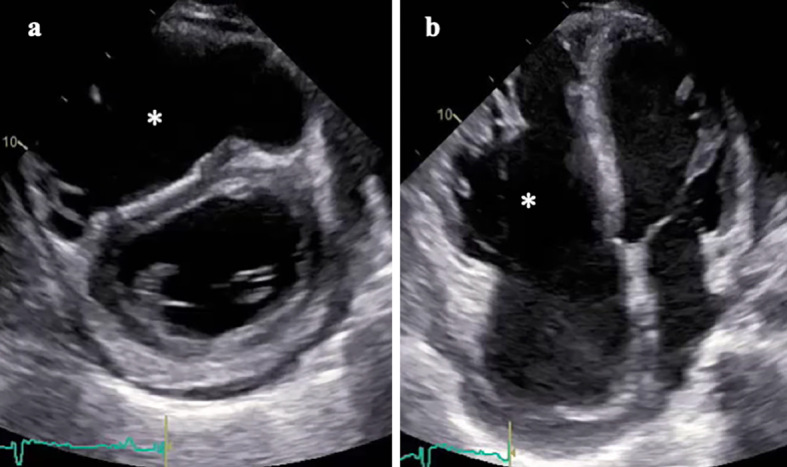
Echocardiography. Parasternal short axis (a) and apical four-chamber images (b) exhibiting right ventricular dilatation and dysfunction (asterisk) with flattened ventricular septum. Severe tricuspid regurgitation and left ventricular dilatation/dysfunction were also observed.