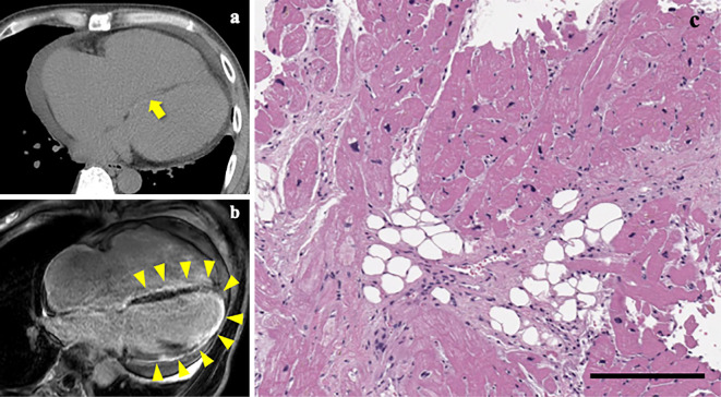
Figure 7.
CT scans, MR images and endomyocardial biopsy specimens. CT scans (a) and MR images (b) depicting fatty infiltration (arrow) and fibrosis (arrowheads) of the ventricular septum, respectively. Fibrosis of the left ventricle was also identified on MR images (b). Light microscopic examination of the right ventricle tissues demonstrating extensive fibro-fatty replacement of the cardiac myocytes (c). Scale bar, 200 μm.

