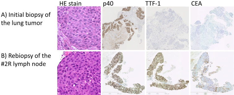
Figure 1.
Biopsy specimens. A) The initial biopsy of the lung tumor. Hematoxylin and Eosin (H&E) staining shows squamous cell carcinoma morphology. Immunostaining with p40 and Cytokeratin 14 (CK14) were positive, while adenocarcinoma markers thyroid transcription factor 1 (TTF-1), carcinoembryonic antigen (CEA), and Napsin A were negative. B) The rebiopsy of the #2R lymph node. H&E staining shows definite squamous cell carcinoma morphology, identical to that of the primary lung tumor. Immunostaining shows features of squamous cell carcinoma (positive p40 and CK 14) along with weak positivity in TTF-1, Napsin A, and CEA.

