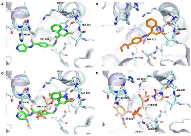
Figure 3.
Comparison of 6b (A,C) and UDP-5S-GlcNAc (C,D) binding mode in the OGT binding site (PDB entry: 4GYY); predicted binding pose for 3b (B). The ligand and the neighboring protein side-chains are shown as stick models, colored according to the chemical atom type (blue, N; red, O; orange, S; green, Cl). Hydrogen bonds are indicated by black dotted lines. Thr922 is doubled due to static disorder.

